Content by: Tanvi Achanti, Chandni
Design by: Prakhya Gupta, Bhavya Jain

THE RAMSTEDT OPERATION FOR PYLORIC STENOSIS
The Ramstedt operation, a surgical procedure revolutionizing the treatment of pyloric stenosis in infants, is one of the most intriguing discoveries in all of pediatric surgery.
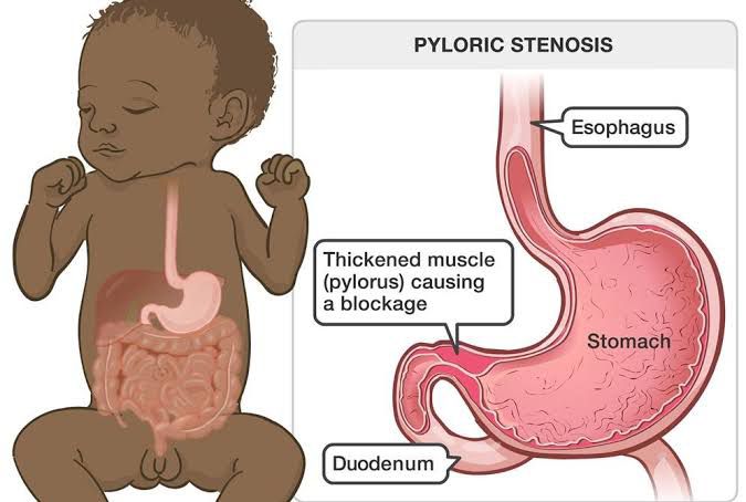
Pyloric stenosis was then considered a life threatening condition, characterized by narrowing of the passage between the stomach and the small intestine due to hypertrophy of the circular muscle of pylorus presenting in severe cases of dehydration, vomiting and weight loss in the young infants barely days old. It was at this time, in 1912 that a German surgeon Conrad Ramstedt was confronted with a case of the same. Before 1912, treatments included gastroenterostomy, pyloroplasty and forcible dilation etc. So in attempts to perform a gastroenterostomy to fix the issue, Ramstedt makes a mishap that helps many many future generations to come.
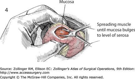
As he was dissecting the thickened pylorus muscle, his scalpel slips from his hand and makes a slight incision on the muscle. Contrary to what could have been a life endangering incident, this laceration results in the immediate alleviation of the obstruction, restoring normal gastric functions.
Rejuvenated by this discovery, he sets up documenting the case and researching further questions that arose. Through repetitive clinical cases and experiments, he refined the technique (now known as Ramstedt pyloromytomy) ultimately finding a standard surgical core for pyloric stenosis, now used in hospitals all around the world now.
As we learn about the story of the Ramstedt operation, the hydrocephalus cure and the balloon dilator and the discovery of it all, we ought to remember that innovation often lies not in perfection or unblemished experiments but in the willingness to embrace the unexpected, to question convention, and to persist in the face of adversity
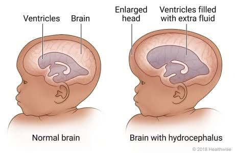
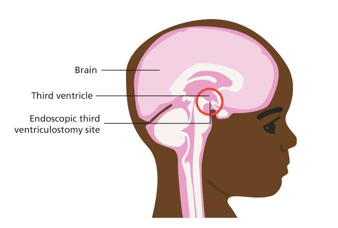
ENDOSCOPIC THIRD VENTRICULOSTOMY FOR SURGICAL CORRECTION OF HYDROCEPHALOUS
The story of the EVT procedure’s accidental discovery is quite fascinating. In the late 1970s, a group of doctors was working on developing new catheters for diagnostic purposes, aiming to visualize the inside of blood vessels more clearly. During one of their experiments, they encountered an unexpected result: they found that by maneuvering the catheter in certain ways, they could actually treat blockages and abnormalities within the blood vessels themselves.
Initially, this discovery was met with skepticism and disbelief. However, as they continued to experiment and refine their techniques, the doctors realized the potential of what they had stumbled upon. They began to explore the possibilities of using these catheters for therapeutic purposes, rather than just diagnostic ones.

Through years of trial and error, along with continuous advancements in technology and medical knowledge, the EVT procedure gradually evolved into a highly effective method for treating a wide range of vascular conditions. Today, it is considered a cornerstone of interventional medicine, saving countless lives and improving outcomes for patients around the world. What started as an accidental discovery has transformed into a vital tool in the fight against vascular disease
.
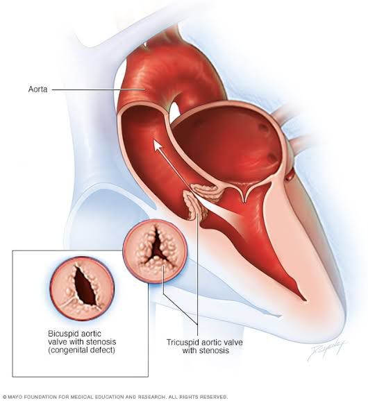
USAGE OF BALLOON CATHETER IN THE TREATMENT OF AORTIC STENOSIS
The accidental discovery of using a balloon catheter to treat aortic stenosis in infants is an intriguing story that highlights the serendipitous nature of medical innovation. In the early 1980s, Dr. William Rashkind, a paediatric cardiologist at the Children’s Hospital of Philadelphia, was treating a newborn with critical aortic stenosis, a condition where the aortic valve is narrowed, obstructing blood flow from the heart to the body.
During a cardiac catheterization procedure intended to assess the severity of the infant’s condition, Dr. Rashkind encountered an unexpected complication: the catheter accidentally lodged across the aortic valve. However, instead of causing harm, this unintended placement had a remarkable effect. When contrast dye was injected through the catheter, it inflated like a balloon, temporarily dilating the narrowed aortic valve and improving blood flow.

Recognizing the potential of this accidental discovery, Dr. Rashkind and his colleague, Dr. William O. Norwood, further explored the use of balloon catheters to treat aortic stenosis in infants. They developed a minimally invasive procedure called balloon valvuloplasty, which involved inserting a balloon-tipped catheter into the narrowed valve and inflating it to widen the opening.
This groundbreaking technique revolutionized the treatment of critical aortic stenosis in infants, offering a less invasive alternative to traditional surgical interventions. Balloon valvuloplasty quickly gained acceptance and became a standard procedure in paediatric cardiology, saving countless lives and improving outcomes for infants with congenital heart defects.